October 2017
POST
DOC POSITION OPEN
OLIGOMERIZATION STATE OF GPCRs AT THE SURFACE OF LIVING CELLS AS EXPLORED BY ATOMIC FORCE MICROSCOPY
A 33 months post-doctoral position (ANR french funding “GRApHICS”) is available for a highly motivated post-doc fellow to join a research group specialized in the pharmacology of G Protein Coupled Receptor (GPCR) to study the cell surface organization of these receptors in living cells using AFM- single molecule force spectroscopy.
October 2017
Paper accepted in Frontiers in Microbiology
Integration of Biochemical, Biophysical and Transcriptomics Data for Investigating the Structural and Nanomechanical Properties of the Yeast Cell Wall
 Marion
Schiavone1,2,
Marion
Schiavone1,2,  Sébastien
Déjean3, Nathalie
Sieczkowski2, Mathieu
Castex2, Etienne
Dague4 and Jean
M. François1*
Sébastien
Déjean3, Nathalie
Sieczkowski2, Mathieu
Castex2, Etienne
Dague4 and Jean
M. François1*- 1Laboratoire d'Ingénierie des Systèmes Biologiques et Procédés, Institut National des Sciences Appliquées de Toulouse, UPS, INP, Université de Toulouse, Toulouse, France
- 2Lallemand SAS, Blagnac, France
- 3Institut de Mathématiques de Toulouse, Toulouse, France
- 4Laboratoire D'analyse et D'architecture des Systèmes du-Centre National de la Recherche Scientifique, Université de Toulouse, Toulouse, France
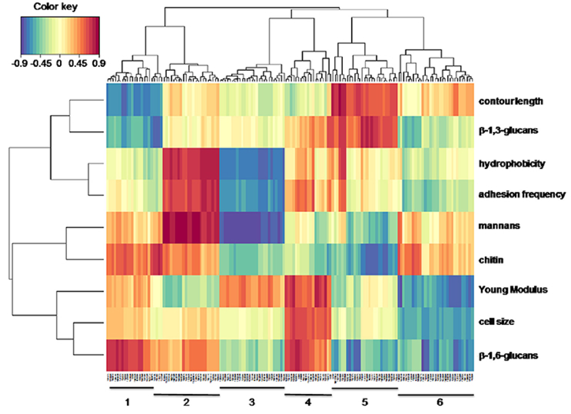
Heat map representation of association between biophysical-biochemical variables and genes transcripts from the laboratory and industrial strains. Biophysical and biochemical variables are presented on the vertical scale whereas the gene transcripts are on the horizontal scale. Clustering analysis made with the mixOmics tool (http://mixomics.org/) allows grouping biochemical and biophysical variables into 4 groups (horizontal) and into 6 groups with respect to gene transcripts (vertical). Red (blue) indicates high positive (negative) correlation between gene transcript and biochemical/biophysical variables.
Most Cited Micron Articles
Ranked #7:
September 2017
Accepted paper in Brain Structure and Function
Nanoscale structural mapping as a measure of maturation in the
murine frontal cortex
G. Smolyakov E. Dague C. Roux M. H. Seguelas C. Gales J. M. Senard D. N. Arvanitis
Excellent collaboration between I2MC and LAAS-CNRS. Application of AFM technology to explore the brain nanostructure. We used both topographical and mechanical mapping to explore the modification occuring during maturation in the frontal cortex.

Atomic force microscopy (AFM) is emerging as an innovative tool to phenotype the brain. This study
demonstrates the utility of AFM to determine nanomechanical and nanostructural features of the murine dorsolateral frontal cortex from weaning to adulthood. We found an increase in tissue stiffness of the primary somatosensory cortex with age, along with an increased cortical mechanical heterogeneity. To characterize the features potentially responsible for this heterogeneity, we applied AFM scan mode to directly image the topography of thin sections of the primary somatosensory cortical layers II/III, IV and V/VI. Topographical mapping of the cortical layers at successive ages showed progressive smoothing of the surface. Topographical images were also compared with histochemically derived morphological information, which demonstrated the deposition of perineuronal nets, important extracellular components and markers of maturity. Our work demonstrates that high-resolution AFM images can be used to determine the nanostructural properties of cortical maturation, well beyond embryonic and postnatal development. Furthermore, it may offer a new method for brain phenotyping and screening to uncover topographical changes in early stages of neurodegenerative diseases.
invited talk and Tutorial
at IMRC 2017, Mexico, 19-26 september 2017
tutorial: Atomic Force Microscopy in Biology: Tricks and solutions
Lecture: Biomedical applications of AFM
June 2017
ANR Project granted: GRAPHICs
Probing GPCR architecture on living cells using AFM-single molecule force spectroscopy
Collaborative Project with Céline Gales, I2MC, Emmanuelle Trévisiol, LAAS-CNR and Jean-Marc Azais, Institut de Mathématique de Toulouse
3 years Post-Doc position opens in january 2018

Invited review in
Seminars in Cell and Developmental Biology
Cell biology of microbes and pharmacology of antimicrobial drugs explored by Atomic Force Microscopy
Cécile Formosa-Daguea, b, c, , 1, Raphaël Emmanuel Duvalb, c, d, Etienne Daguea,
a
LAAS-CNRS,
Université de Toulouse, CNRS, Toulouse, France
b
CNRS,
UMR 7565, SRSMC, F-54506 Vandœuvre-lès-Nancy, France
c
Université
de Lorraine, UMR 7565, SRSMC, Faculté de Pharmacie,
F-54001 Nancy, France d
ABC
Platform®,
F-54001 Nancy, France

Nanomechanical analysis of the capsule of Klebsiella pneumoniae. (a) Mechanical model for cell indentation of encapsulated bacteria by an AFM tip proposed by Wang et al. In this model, four stages are identified. Capsule polysaccharides are the green lines and the red lines represent fimbriae. Each stage corresponds to a particular regime of the force profile presented on the graph. Stage 1: long-range double layer repulsion between the negatively charged bacterial surface and the negatively charged silicon nitride AFM tip, fitted to double layer theory (green line); Stage 2: steric or electrosteric interaction during compression of cell surface polymers, fitted to the Pincus theory (purple line); Stage 3: elastic deformation of the compacted surface polymers, fitted to the Hertz theory (blue line); and Stage 4: compression of the bacterial cell cytoplasm, fitted to Hooke's law (black line). (b) Indentation curves analysis of Formosa et al. reveals the capsular organization of colistin-resistant K. pneumoniae cells. The inset represent a force curve recorded on a cell in native condition, fitted to the Hertz model. (c) Mechanical model of K. pneumoniae cell indentation proposed by Mularski et al. In this model, first stage consists in long-range double layer interaction between charged tip and surface, then the tip indents the capsular polysaccharide and finally the cytoplasm. The graph represents a typical force profile of K. pneumoniae cell in HEPES buffer (red) with Hooke’s law fit (blue) and derived parameters, bacterial spring constant, Kbacterium = 97 mN/m, and capsule thickness = 340 nm. Reprinted with per- mission from Refs. [35–37].
March 2017
Accepted paper in Journal of Structural Biology
Biophysical properties of cardiomyocyte surface
explored by multiparametric AFM
Congratulations to Georges for his great work on this topic
This publication is the result of a great collaboration between, LAAS and ITAV-CNRS, I2MC-INSERM.

Elasticity map of a cardiomyocyte recorded in AFM QNM mode (on the left) and its related analysis by fourrier transformation (on the right) demonstrating the structural periodicity.
January 2017
Campus France, chargé de la gestion des Partenariats Hubert Curien (PHC), pour le compte du ministère des Affaires étrangères et du développement international, a le plaisir de vous faire savoir que le Comité mixte du Programme AURORA a retenu votre projet au titre de l'exercice 2017.Elucidating
interaction
capacities of microorganisms using sensitive force probes
`

December 2016
Excellent stay at ITN Mexico for Automatip project
We are really doing good job together. Excellent collaboration
November 2016
We were pleased and proud to receive Adrian Martinez, Karen Genesis and Sergio Proa from Institute Polytechnic of Mexico
to collaborate on our Automatip Project. The Mexican-French exchange is funded by ECOS Nord Committee.

2016 november the 24th
Congratulations to Cécile who received the DGA PhD award. The award has been given during "Forum de l'innovation DGA" by Jean-Yves Le Drian, ministre de la Défense.
Cécile Formosa, thèse LAAS (Laboratoire d'analyse et architecture des systèmes) au CNRS Toulouse, pour ses travaux visant à « comprendre les cibles d’action des molécules antimicrobiennes à l’aide des nanotechnologies ». Il s’agit d’évaluer l’action de nouvelles molécules antimicrobiennes (antibiotiques et
antifongiques) en utilisant des techniques innovantes de microscopie à force atomique (AFM). L’objectif est de lutter contre l’augmentation de la résistance des bactéries aux antibiotiques observée à l’échelle mondiale, une menace pour la santé publique ainsi que celle des militaires en opérations extérieures.

2016 october the 11th
PhD Defense: Véronique Lachaize.

Véronique brillantly defended her PhD thesis.
Congratulations
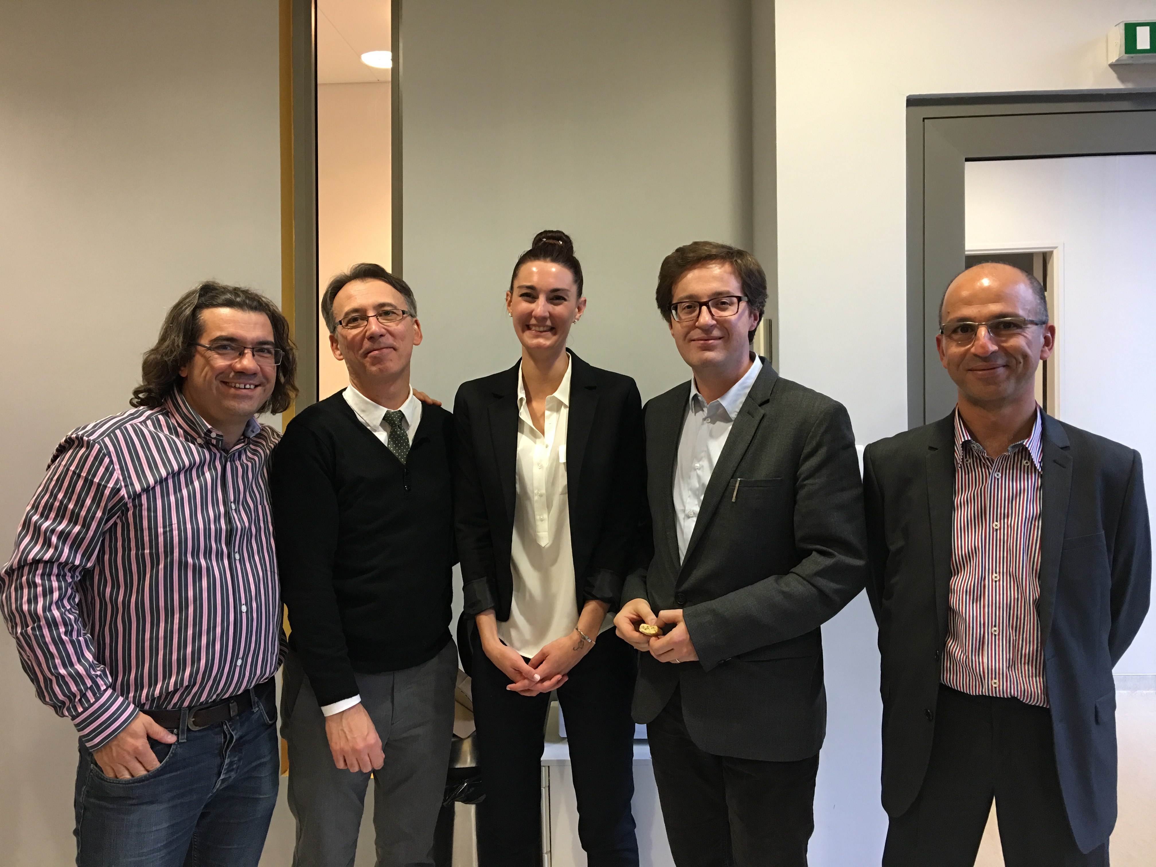
from the left to the right:
Childérick Severac, Philippe Manivet, Véronique Lachaize, Etienne Dague and Sid Labdi. Sofiane El Kirat-Chatel, the last committy member is missing on the picture.
October 2016
Accepted paper in
ACS applied materials and interfaces
Georges Smolyakov,a,b Bénédicte Thiebot,c Clément Campillo*,c Sid Labdi,c Childerick Severac,a Juan Pelta,*c Étienne Dague*a,b
aITAV CNRS, Université de Toulouse, CNRS, UPS, France b LAAS-CNRS, Université de Toulouse, CNRS, Toulouse, France
cLaboratoire d’Analyse et Modélisation pour la Biologie et l'Environnement LAMBE-CNRS, Université d’Evry,Evry, Université de Cergy-Pontoise, Cergy-Pontoise,
France
This publication is the result of a great collaboration between, LAAS and ITAV-CNRS, University of Evry and University of Cergy. Let the show go on and continue this exciting work.
September 2016
Book Chapter in
Handbook of Electroporation,
edited by Damijan Miklavčič
Atomic Force Microscopy to explore
Electroporation effects on cells
In this chapter, we review the work of Louise Chopinet, who graduated in 2013, and Flavien Pillet who is currently on a post doc position at IPBS. Many thanks for their contributions, this chapter is dedicated to their work.
http://link.springer.com/referenceworkentry/10.1007/978-3-319-26779-1_134-1
July 2016
New name for our team at LAAS-CNRS:
ELIA Engineering for Life Science Application

June 2016
Organization of Nano In BIO


NanoInBio
Advances for life and materials science (LeGosier, Guadeloupe,
France, 31 mai – 5 juin 2106)
-
Hélène Martin-Yken H., Formosa-Dague C., Schiavone M.,
François J-M., Dague E., Stress, Drug Resistance and
Adhesion: a closer look into the dark side of the wall
-
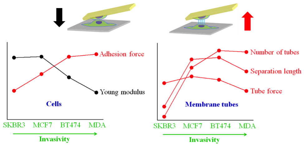
Smolyakov G., Campillo C., Labdi S., Thiebot B., Severac C.,
Pelta J., Dague E. Cell adhesion and rigidity probed
by single-cell force spectroscopy reveal the invasive
character of breast cancer lines
-
Schiavone M., François J-M., Sletmoen M., Dague E.,
Interactions between concanavalin A and yeast cell wall
mutants studied by Optical Tweezers and Atomic Force
microscopy
-
Pillet F., Formosa-Dague C., Teissié J., Rols M-P., Dague
E. AFM analysis of cell-wall damages induced on bacteria
by Pulsed Electric Field
May 2016
Invited speaker at Imaging the Yeast Symposium
Dague E., Imaging living yeast cells and quantifying their biophysical properties by atomic force microscopy
April 2016
Opened PhD position in the team
NANOCARD II
Caractérisation de la membrane latérale du cardiomyocyte à l’échelle nanométrique en physio-physiopathologie cardiaque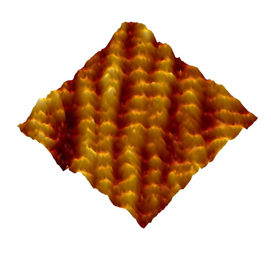
March 2016: France-Mexico Cooperation
ECOS Nord Project awarded to Etienne, Childérick and Adrian
Nano-palpation pour le diagnostic
- France : L'Institut des Technologies Avancées en sciences du Vivant
(ITAV) du Centre National de la Recherche
scientifique (CNRS)
-
Mexique : Centre
de recherche en informatique (CIC) MICROSE lab de l’Institut
Polytechnique National (Mexico)
February 2016: IDEX Project awarded to Hélene, Mickael, Céline, Marit and Etienne
SMiRCH:
Force Spectroscopy and Optical Tweezers combined for Single Molecule Research at the Surface of Living Cells

January 2016: coopération France-Norvège
Candidature Åsgard Acceptée
Mon séjour en Norvège est prévu pour octobre 2016!
Le programme Åsgard a pour objectifs de:
- Développer les échanges scientifiques entre la France et la Norvège
- Favoriser l’émergence de coopérations en recherche et en transfert de technologie
- Permettre aux chercheurs français et norvégiens d’étendre leurs réseaux internationaux
November 2015
- Congratulation to Laure and Flavien for their poster award received during 11ème journée du Canceropole Grand Sud Ouest
And
- Invited keynote lecture at NanoBioMed in Spain
11ème journée du cancéropole Grand Sud Ouest (Bordeaux, France, 5-6 novembre 2015)
Pillet F., Gibot L., Madi M., Rols P-P., Dague E., Atomic Force Microscopy: a tool to study native dermis and 3D cellular models. NanoBioMed 2015 (Barcelone, Spain, 18-20 nov 2015) Dague E. Biomedical Applications of Atomic Force Microscopy

juillet 2015
Congratulation to Cécile for her PhD award given by "Société Française des microscopistes"
XIVème colloque de la Société Française
des Microscopies (Nice, France 30 juin-3 juillet 2015)
Formosa
C., Dague E., Comprehension of the mechanisms of actions of
antimicrobial drugs using bionantotechnologies
(invited
plenary lecture)
March 2015
Chapter published in the book Advanced Microscopy in mycology
. Dague E., Formosa C., 2015, Imaging living yeasts cells and quantifying their biophysical properties by Atomic Force Microscopy in Springer Eds, Tanya Dahms and Kirk Czymmek

February 2015
3 papers published in
- FEMS Yeast Reserach: congratulations to Marion
- Journal of Molecular Recognition: congratulations to Cécile and Véronqiue
- Nanomedicine NBM: congratulations to Cécile
Formosa C., Lachaize V., Rols M.P., Martin Yken H., François J.M., Duval R.E., Dague E., 2015. Mapping HA-tagged protein at the surface of living cells by atomic force microscopy.
Journal of Molecular Recognition 28. 1-9
Formosa C., Schiavone M., Boisramé A., Lavie Richard M., Duval R.E., Dague E., 2015. Multiparametric imaging of adhesive nanodomains at the surface of C. albicans by Atomic Force Microscopy
Nanomedicine NBM
january 2015
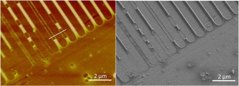
New paper published in Nature Protocols
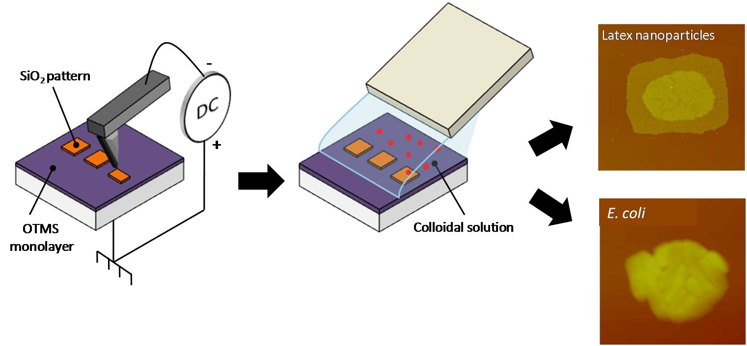
Formosa C., Pillet F., Schiavone M., Duval R.E., Ressier L., Dague E., 2015.
Generation of living cell arrays for Atomic Force Microscopy studies
Nature Protocols 10. 1. 199-204

December 2014 :
- Marion Schiavone PhD Thesis
Combination of biochemical, molecular and biophysical approaches to investigate the impact of strain background and production process on the yeast cell wall composition and molecular architecture- new publication in ACS Applied materials and interfaces
- Oral and Poster presentation at AFMBioMed, SanDiego California USA

Congratulation to Marion Schiavone for the defense of her PhD thesis. She will be hired on a Post Doc position soon by Lallemand SA.
Jauvert E., Palleau E., Dague E., Ressier L.,
Directed assembly of living Pseudomonas aeruginosa on PEI patterns fabricated by nanoxerography for statistical AFM bio-experiments
ACS Applied materials and interfaces
- Dague E., Three biomedical applications of atomic force microscopy, (Communication orale).
- Formosa C., Duval R.E., Dague E., Unravelling of a mechanism of resistance to colistin in Klebsiella pneumoniae thanks to Atomic Force Microscopy, (Communication orale).
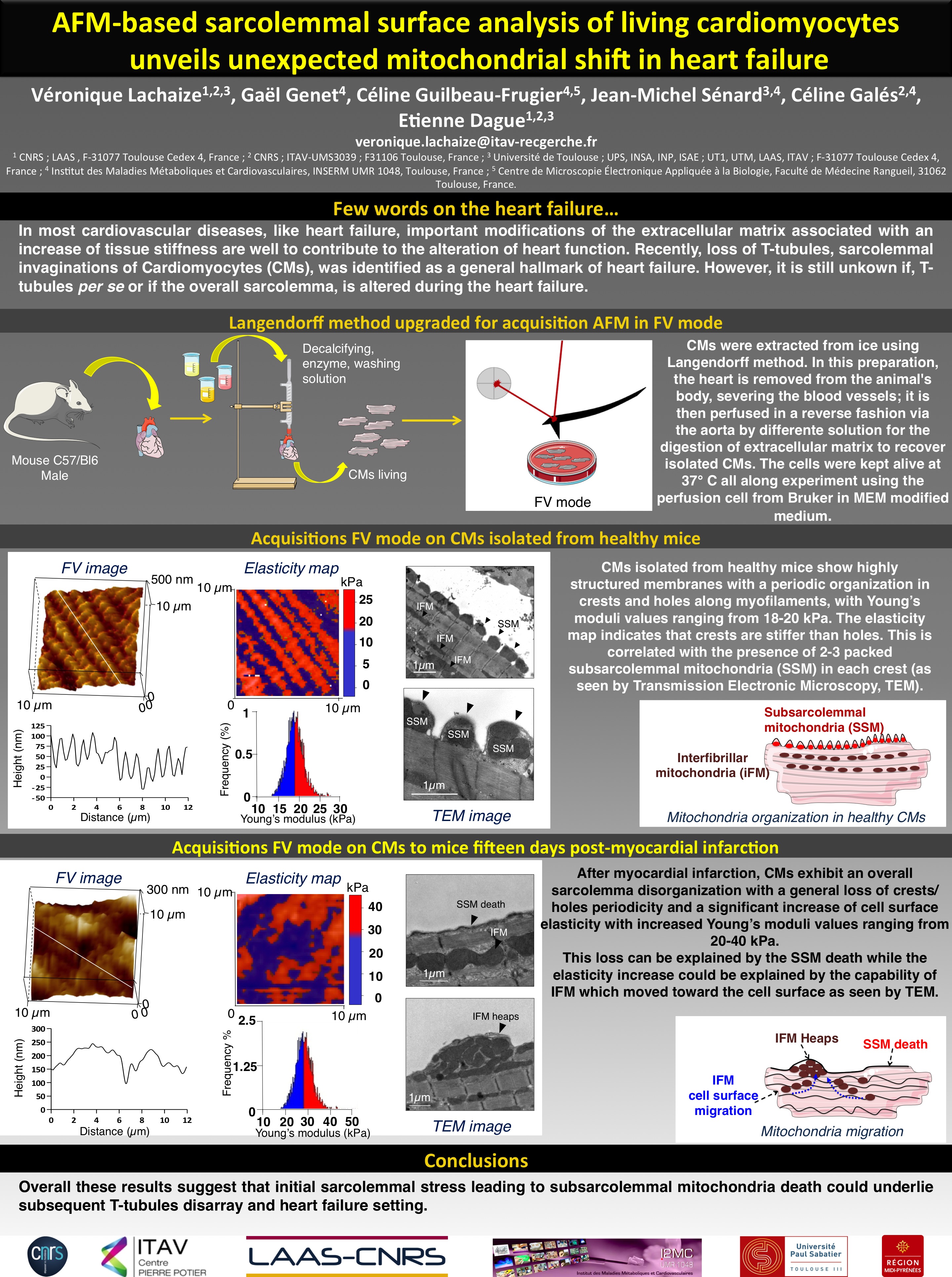
- Lachaize V ;, Genet G., Guilbeau-Frugier C., Severac C., Senard J-M., Gales C., Dague E. AFM-based sarcolemmal surface analysis of living cardiomyocytes unveils unexpected mitochondrial shift in heart failure. (Communication affichée)
September 2014, Cecile Formosa PhD thesis
Comprehension of the mechanisms of action of antimicrobial molecules using nanobiotechnologies
Cécile will stay with us, on a Post-Doc position, for 2 more months.
In Januray she will move on a Post Doc position in the Dufrêne's lab.

July 2014
new Papers accepted in
FEMS YEAST:
A combined chemical and enzymatic method to determine quantitatively the polysaccharaide components in the cell wall of yeasts
by Schiavone M., Formosa C., Martin Yken H., Dague E., François J-M.
Journal of Molecular Recognition
Mapping HA-tagged protein at the surface of living cells by atomic force microscopy.
by Formosa C., Lachaize V., Rols M.P., Martin Yken H., François J.M., Duval R.E., Dague E
and Nanomedicine NBM
Multiparametric imaging of adhesive nanodomains at the surface of C. albicans by Atomic Force Microscopy
by Formosa C., Schiavone M., Boisramé A., Lavie Richard M., Duval R.E., Dague E.
June 2014
Welcome to Georgiy Smolyakov. He joined the group at ITAv and is funded by a grant from "Fondation pour la Recherche Médicale" to work on the Nanocardiology project.
This research is supported by FRM, Grant # ING 21040129094
MAY 2014
New paper published in Journal of Molecular and Cellular Cardiology
AFM of living cardiomyocytes surface unveiled unexpected mitochondrial shifts in heart failure
by
Dague
E., Genet G., Fauconnier J., Guilbeau-Frugier C., Payré B.,
Chopinet L., Alsteens D., Severac C., Thireau J., Heymes C.,
Honton B., Lacampagne A.,Pathak A., Sénard J.M., Galés C.
Forum des microscopistes à sonde locale
17-21 mars 2014 Montauban

February 2014
Oral and poster presentations at the linz winterworkshop


Position Available for 2 years
Engineer with excellent skills in AFM applied to living cells
Funded by FRM (Fondation pour la Recherche Médicale), the project aims at exploring the cardiomyocytes organization at the nanoscale using atomic force microscopy. For more details contact
edague@laas.fr and celine.gales@inserm.fr
January 2014 Paper accepted in BMC Biology
Uncovering by AFM an original circular structure at the yeast cell surface in response to heat shock
by F. Pillet, S. Lemonier, M. Schiavone, C. Formosa, H. Martin-Yken, J-M François, E Dague

January 2014
HAPPY NEW YEAR
December 2013
New paper accepted in BBA General Subject

21 novembre 2013
Poster presentation at Forum de l'Innovation of DGA (Direction Générale de l'Armement).
Bionanotechnologies : Comprendre les cibles d'action de nouvelles molécules antibactériennes
Thèse de Cécile Formosa
La direction générale de l’armement (DGA) a organisé son second forum innovation le 21 novembre 2013 sur le campus de l’école polytechnique à Palaiseau. Articulée autour d’un village exposition, d’une séance plénière et d’ateliers thématiques, cette manifestation est l'événement annuel majeur de la recherche et de l’innovation duale.
Novembre 2013
Invited lecture in Tronheim, Norway

Intensive course for PhD students from all over Norway within the fields Biophysics, Biotechnolody, Pharmacy, Medicine or related fields. They will gather in Trondheim on these two weeks. Week one they learn about light microscopy based techniques, and week two, which is the week starting with the 11th of November, they learn about force based techniques, involving AFM and optical tweezers.
Dague E., From single cell to single molecule, AFM in life sciences
Octobre 2013
Bienvenue à Véronique Lachaize
Doctorante co encadrée avec Céline Gales de l'I2MC,
membre de l'équipe projet NanoCardiologie à l'ITAV USR 3505
October 2013
New publication in Current genetics

September 2013
New publication in PlosOne

24 septembre 2013
Soutenance Thèse Louise Chopinet
Directeurs de ThèseMarie Pierre Rols, Etienne Dague
Rapporteurs :
Luis Mir, Victor Shahin
Examinateurs :
Bernard Ducommun, Philippe Pouliguen, René Vézinet

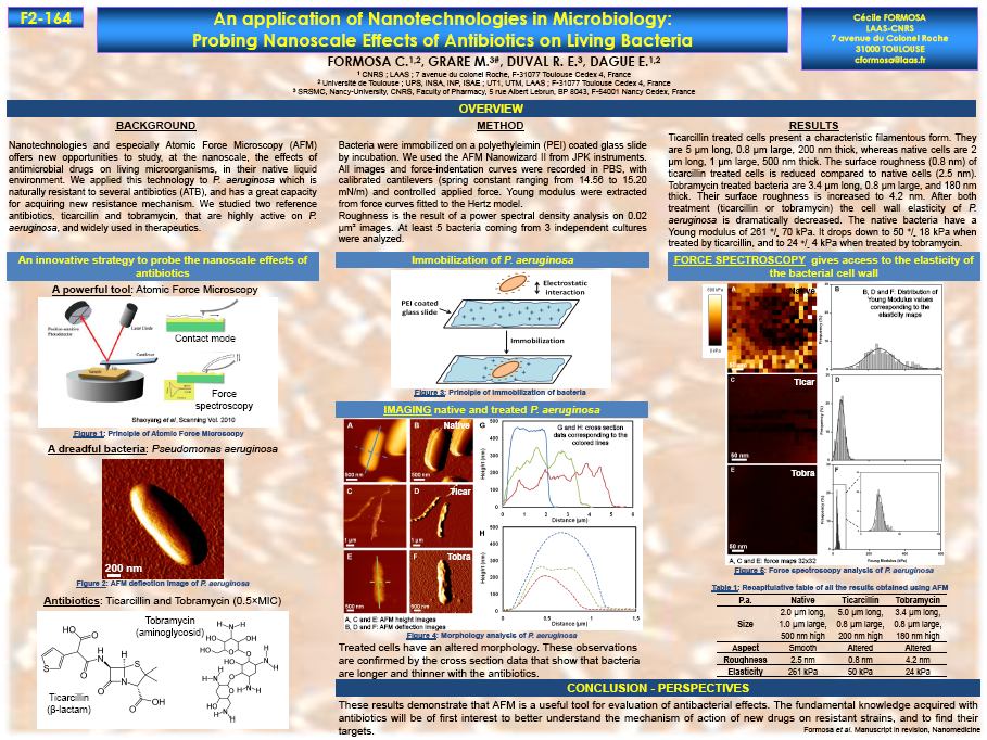
ICAAC 2013 in Denver
Etienne was honored as George
McCracken Infectious Disease Fellow and received a grant to
cover travel expenses
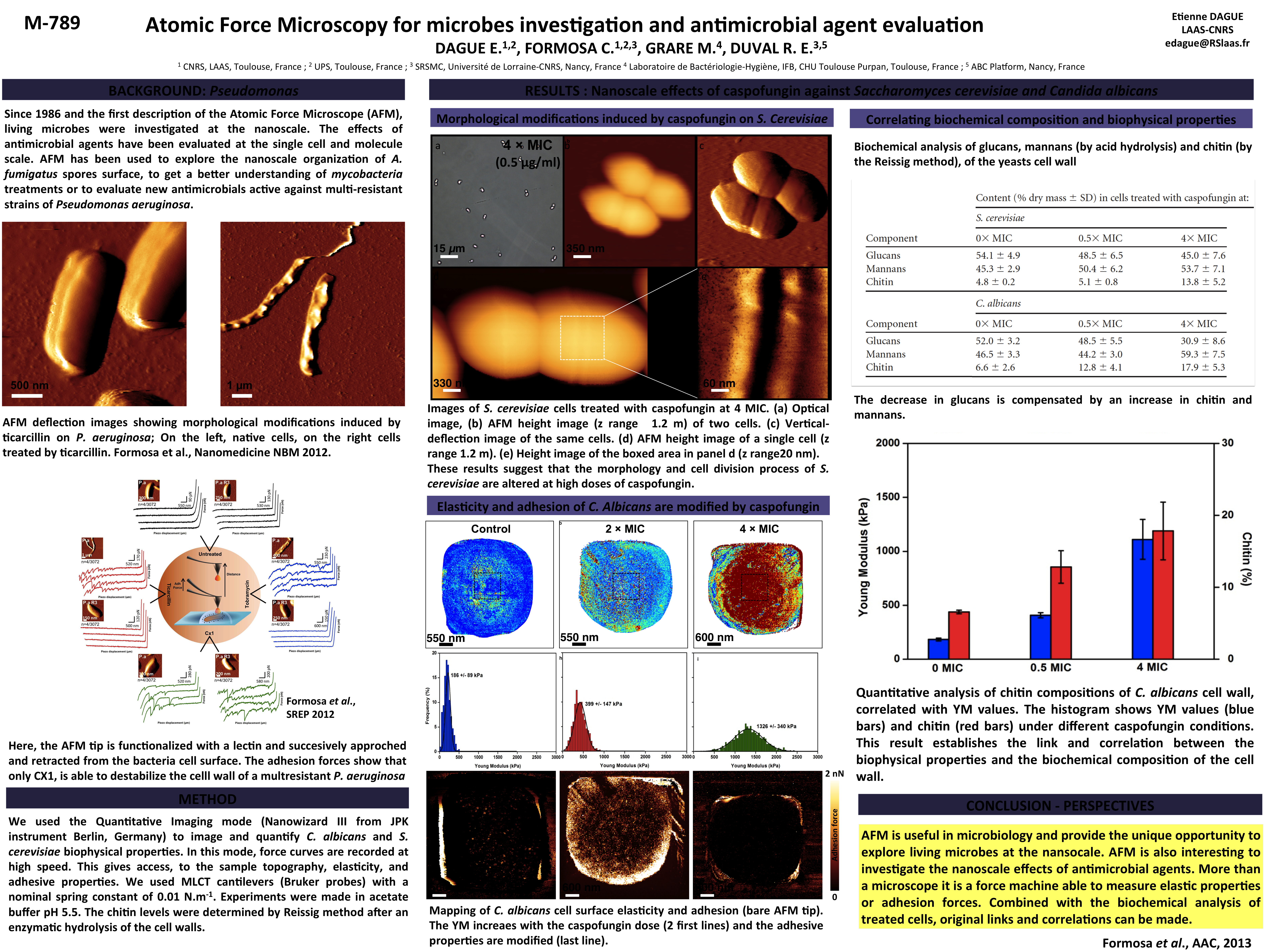
Dague E., Formosa C., Grare M., Duval R.E., Atomic
Force Microscopy for microbes investigation and antimicrobial
agent evaluation
ICAAC 54th Interscience conference on antimicrobial agents and chemotherapy (Denver, USA, 10-13 septembre 2013
![]()
5 juillet 2013
Projet Région Bacteau retenu pour financement par la région Midi Pyrénée
Le travail sera réalisé par Arthur Gaveau, doctorant, et pour ce qui me concerne sera hébergé sur la plate forme de BioNanoTechnologie de l'ITAV USR 3505
Poster communication at PYFF5
5th Conference on Physiology of Yeast and Filamentous Fungi
June 4 - 7, 2013, Montpellier, FRANCE

The yeast response to
external stress investigated by Atomic Force Microscopy
C.
Formosa, M. Schiavone, C. Elsztein, H. Martin-Yken, Jr M. A.
de Morais, R. E. Duval, J. M. François and E.
Dague
and
A
simple method to determine Saccharomyces cerevisiae cell wall
composition by combined chemical and enzymatic hydrolysis
M.
Schiavone, J.-M. François
June 2013
New publication accepted in
Biophysica and Biochimica Acta; Biomembranes

Destabilization
induced
by electropermeabilization analyzed by Atomic Force Microscopy
Louise Chopinet, Charles Roduit , Marie-Pierre Rols and Etienne Dague
Highlights
We demonstrate that AFM can be used to study electroporation.
Electric fields provoke a decrease in membrane stiffness.
Electro-destabilization process measured by AFM happens later than
membrane permeabilization
Electro-destabilization last longer than PI uptake observed by
fluorescence imaging.
AFM imaging shows the disturbed plasma membrane.
May 23rd 2013
AFM Workshop at Institut du Cerveau et de la Moelle épinière
(Paris)
Organized for INSERM by Etienne Dague and Grégory Francius.
Program
Cellular and molecular nanomechanics by Atomic Force
Microscopy
23 mai 2013 - May 23rd, 2013
Organizers: Etienne Dague (LAAS-CNRS UPR 8001, Toulouse,France) et Grégory Francius (LCPME UMR 7564, Nancy, France)
May 16th 2013
L'AFM dans tous ses états
Rencontre de la fédération de recherche FERMaT

April 2013
New publication in Antimicrobial Agent and Chemotherapy
Nanoscale effects of Caspofungin against two yeast
species; Saccharomyces
cerevisiae and Candida
albicans
C. Formosa, M. Schiavone, H. Martin-Yken, J. M. François, R. E. Duval and E. Dague
LAAS-CNRS, LISBP, SRSMC, Plateforme ABS
In this paper, the authors charaterize the effect of caspofungin against S. cerevisiae and C. albicans. The use of AFM for investigating the effect of antifungal drug is relevant in nanomedicine, as it should help understanding their mechanism of action on the fungal cells, as well as unraveling unexpected effects on cell division and fungal adhesion.

7 février 2013
Publication acceptée dans MICRON
Imaging
living
cells surface and quantifying its properties at high
resolution using AFM in QITM mode
Chopinet, L. and Formosa, C. Rols, M. P. Duval, R. E.5 Dague, E.
This paper presents results obtained using the quantitative imaging mode. We explored the surface topography, nanomechanical or adhesion properties of several single cells from bacteria to eukaryotic cells :
- Escherichia coli
- Candida albicans
- Aspergillus fumigatus
- CHO cells
- cell nuclei
All together the results demonstrate that AFM can be conducted on soft and fragile sample, very close to their native environment. All the experiments were conducted in liquid, on living cells, at regulated temperature and pH when required.
Soutenance de Thèse : ERIC JAUVERT
SALLE DES THÈSES DE L'INSA
21 DÉCEMBRE À 10H
Thèse co diriger par Laurence Ressier (LPCNO) et Etienne Dague (LAAS-CNRS)
Sujet : Assemblage dirigé de micro organismes et fonctionnalisation de pointes pour biocaractérisation par AFM
November 2012
Habilitation à Diriger les Recherches
Etienne DAGUE
Docteur de l’université Henri Poincaré Nancy 1,
Docteur en Pharmacie,
Chargé de Recherche du CNRS au LAAS-CNRS
(UPR8001)
Sujet
Les
technologies de Microscopie à Force Atomique appliquées sur
les systèmes vivants
Membres du jury
Dr.
Sandor Kasas, EPFL-Univeristé de Lausanne (CH)
Dr.
Frank Lafont, DR CNRS INSERM U1019 - CNRS UMR 8204
Dr.
Pierre-Emmanuel Milhiet, DR CNRS UMR 5048
Examinateurs
Dr.
Jean Pierre Aimé, DR CNRS Université de Bordeaux 1, UMR 5248
Dr.
Christian Bergaud, DR CRNS, LAAS-CNRS UPR 8001
Pr.
Pierre-Emmanuel Gleizes, Université de Toulouse, LBME
UMR 5099
Pr.
Christophe Vieu, INSA Université de Toulouse, LAAS-CNRS UPR
8001
September 2012
Academic Editor of PlosOne

September 2012 :
NANOCARDIOLOGY AND BIOPHYSIC OF CANCEROUS CELLS
NEW TEAM IN CENTRE PIERRE POTIER ITAV
August 2012
New paper in Scientific Reports,
Nature Publishing Group

Nanomedecine NBM Impact Factor 2012
IF = 6.692!!
april 2012
New paper in Sensors and Actuators

Mai 2012
New publication in Biofouling

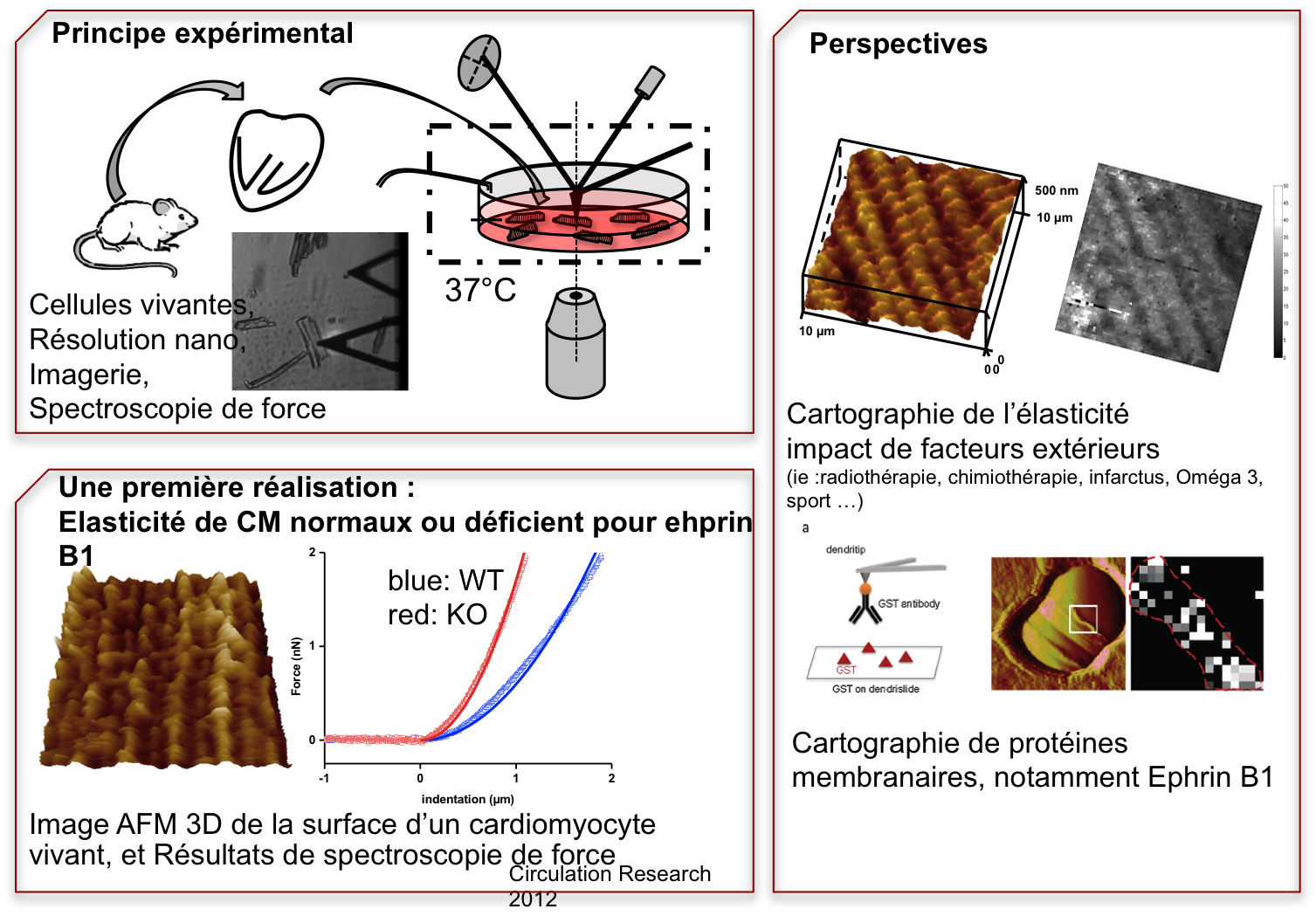
New Publication in Circulation Research
Genet G., Guilbeau-Frugier C., Honton B., Dague E., Schneider M.D., Coatrieux C., Calise D., Cardin C., Nieto C., Payré B., Dubroca C., Marck P., Heymes C., Dubrac A., Avranitis C., Despas F., Altié M-F.; Seguelas M-H., Delisle M-B., Davy A., Senard J-M., Pathak A.
Ephrin
B1
is a novel component of the lateral
membrane of the cardiomyocyte and is
essential for the stability of cardiac
architecture cohesion
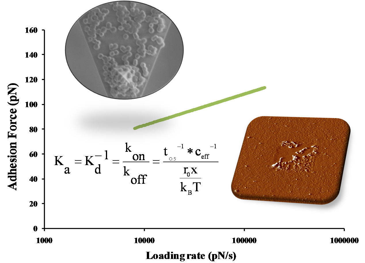
New publication in Biophysical journal
Doan Thanh Lam Le, Yann Guérardel, Pascal Loubière, Muriel Mercier-Bonin and Etienne Dague
Measuring Kinetic Dissociation/Association Constants Between Lactococcus lactis Bacteria and Mucins Using Living Cell Probes
à l'INSA département de mécanique S 112
13h30
Identification des déterminants physico-chimiques et biologiques mis en jeu dans l'adhésion de Lactococcus lactis à la mucine, modèle PGM
10th International Symposium on Scanning probe microscopy and optical tweezers in life sciences
Berlin
AFM applications in nanomedecine, from microbiology to cardiology

From the left to the right (click to enlarge): Paolo Samori (University of Strasbourg), Torsten Jähnke (JPK), Roderick Lim (Basel University), Małgorzata Lekka (Polish Academy of Sciences), Giovanni Longo (EPFL), Nic Mullin (University of Sheffield), Marit Sletmoen (NTNU Trondheim), Etienne Dague (LAAS-CNRS), Bart Hoogenboom (University College London) and Wenke Zhang (Jilin University).
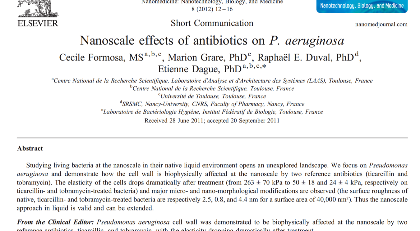
Paper accepted in Nanomedecine NBM
Nanoscale
effects
of antibiotics
on P. aeruginosa

LAAS, ITAV CNRS, Université de Toulouse
SRSMC, UMR CNRS Nancy Université
CHU Purpan
51st interscience conference on antimicrobial agents and chemotherapy
AFMBIOMED Conference

Oral Communication
AFM applications in nanomedecine, from microbiology to cardiology
Paper accepted in Nanotechnology
Assembly of live micro-organisms on microstructured PDMS stamps by convective/capillary deposition for AFM bio-experiments

E. Dague, E. Jauvert, L. Laplatine, B. Viallet,
C. Thibault and L. Ressier
From
LAAS, ITAV, CNRS, Université de Toulouse
LPCNO, INSA, Université de Toulouse
ANR Jeune Chercheur AFMYST
retenu pour financement

Réunion des AFMistes Toulousains
Childérick Severac, ITAV
Etienne Dague, LAAS-CNRS

17 dec 2010
Séminaire Institut de Biologie Structurale (IBS) Grenoble

Cours M2 P : Diagnostic microbiologique : approches innovantes
BioNanotechnologie
LAMBE, Université d'Evry et Cergy Pontoise

Host-Pathogen interactions in respiratory Infectious Diseases
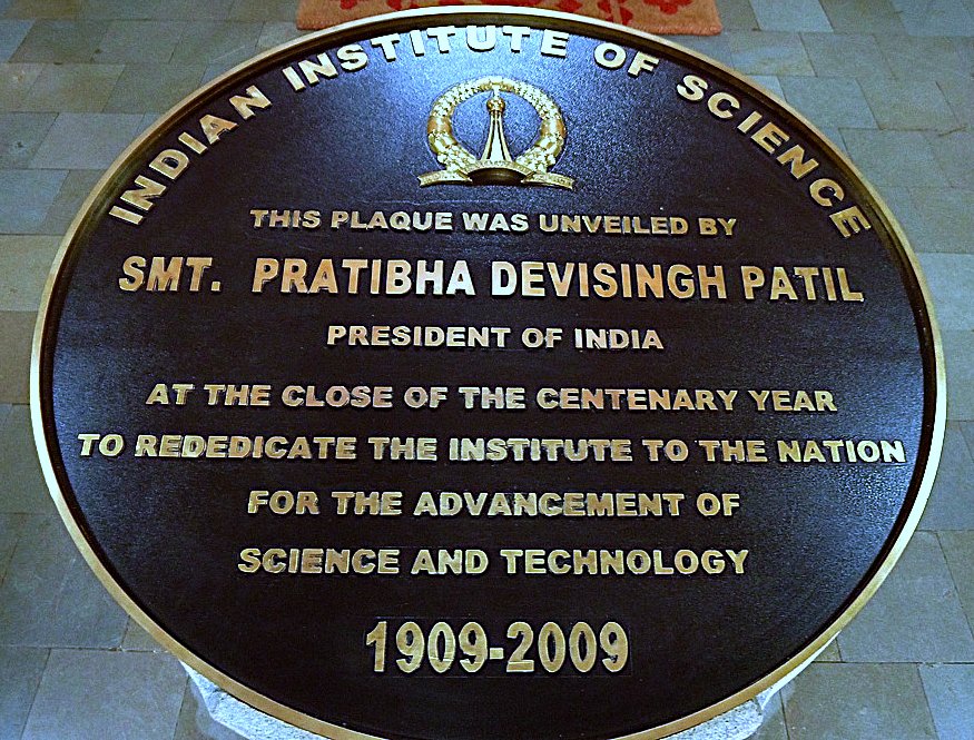
Organized in Bangalore INDIA by CEFIPRA :
Indo-French Centre for the promotion of Advanced Research

Research
Networking
Programme on
The
Functional genomics in Aspergillus fumigatus
and new strategies to fight against the first fungal pathogen
in Europe
(Fuminomics)
Third
Annual Meeting Cell
Biology and Biochemistry
Institut Pasteur, Paris, France
October
7-8, 2010

THERMOBIOAFM
Just do it!

Paper accepted in Langmuir :
Probing
in vitro interactions between
Lactococcus lactis and mucins using AFM

This
paper by
- LISBP - INRA: UMR792 CNRS: UMR5504
-
Laboratoire de Physico-Chimie des Surfaces, CNRS/ENSCP,
UMR7045, ce

The project
“Comparing aptamers and anti-body for single molecule force
spectroscopy experiments” will be funded by the network C’Nano
Grand Sud Ouest. This project, conducted by E. Dague
(LAAS-CNRS), is the result of a budding collaboration with
Emmanuelle Trevisiol from LISPB and Jean-Jacques Toulmé from
IECB. The project aims at developing the aptamère technology in
order to perform single molecule force spectroscopy experiments.
Just do it !
Paper accepted in YEAST


This
paper by
Etienne
Dague,
Rajaa Bittar,
Hubert Ranchon,
Fabien
Durand,
Hélène Martin Yken
and
Jean M François
From
Enjoy
the reading

Découvrez le calendrier des séminaires interne NBS
-
25 mars 2010 : Samuel Guillon
Fabrication
et caractérisation de nanosystèmes électromécaniques à
actionnement et détection piézoélectrique intégré
-
1er avril 2010 :
Thierry Leïchlé
Diélectrophorèse
et nanofentes pour le filtrage et la concentration de
biomolécules
-
8 avril 2009 : Amélie Beduer
La
domestication des neurones, entre fouet et carotte
Nanopatterning molecularly imprinted polymers by soft lithography: a hierarchical approach
Lalo H., Ayela C., Dague E., Haupt K.
Programme ANR jeune chercheur : projet AFMYST
Le projet transdisciplinaire AFMYST se positionne à l’interface de la biologie, de la chimie et de la physique. La question biologique posée concerne l’organisation et la morphogénèse de la paroi des levures Saccharomyces cerevisiae. Cette paroi cellulaire a été essentiellement étudiée par des méthodes de biochimie ou de biologie moléculaire. Notre objectif est d’apporter à cette connaissance les observations en microscopie à force atomique donnant accès à la biophysique de la paroi.

Atelier Interuniversitaire de Microéléctronique
Inauguration du centre Pierre Potier : ITAV

Séminaire annuel du groupe NBS

![]()
Du 30 août au 3 septembre : International conference on molecular mechanisms of fungal cell wall biogenesis
Dague
E.,
Ranchon H., François J-M., β-glucans, mannans and chitin
relative influence on Sacharomyces cerevisiae
cell wall nanomechanical properties (communication affichée)
Nanoscale VII

Using SPM and related techniques
Oral Presentation
2nd ESF/UB European Summer School in Nanomedecine
Quinta da Marinha (Cascais, Lbonne-Portugal)
Communication : E Dague
Nanotechnologies for exploring living cells
Nanowerk spotlights our recent paper in Langmuir entitled Nanomechanical properties of dead or alive single patterned bacteria

| Dead or alive - nanotechnology technique tells the difference | |
| (Nanowerk Spotlight) A major concern in microbiology is to determine whether a bacterium is dead or alive. This crucial question has major consequences in food industry, water supply or health care. While culture-based tests can determine whether bacteria can proliferate and form colonies, these tests are time-consuming and work poorly with certain slow-growing or non-culturable bacteria. They are not suitable for applications where real-time results are needed, e.g. in industrial manufacturing or food processing. | |
| A team of scientists in France has now discovered that living and dead cells can be discriminated with a nanotechnology technique on the basis of their cell wall nanomechanical properties. This finding is totally new and has been made possible thanks to an interdisciplinary approach which mixes physics, biology and chemistry. This work is a key stone in the understanding of bacterial cell wall behavior. |
programme complet des séminaires

LAAS-CNRS club des affiliés, Pole CancerBioSanté
Communication orale : A. Bancaud, E. Dague
Nouvelles méthodes d'imagerie dynamique en 3 dimensions appliquées aux cellules vivantes

Communication Orale, E Dague :
Working on living cells with the Atomic Force Microscope

Langmuir IF = 4.009
Vol. 24 , Issue 23, 13254-13257
Water Research IF = 3.427
vol.42, 4751-4760
Adhesion of Campylobacter jejuni and Mycobacterium avium onto polyethylene terephtalate (PET) used for bottled waters
Josiane-Aurore Tatchou-Nyamsi-König, Etienne Dague, Martine Mullet, Jérôme F.L. Duval, Fabien Gaboriaud, Jean-Claude Block
Vol. 24, Issue 07
April 1, 2008 Cover
Click here to see the article.


Couverture du Journal, The Analyst (IF=3.198) Mars 2008 (lien article diatomées)
Oral communication : Chemical force microscopy of single living cells ; session: Biological AFM application; chaired by Christian LeGrimmellec
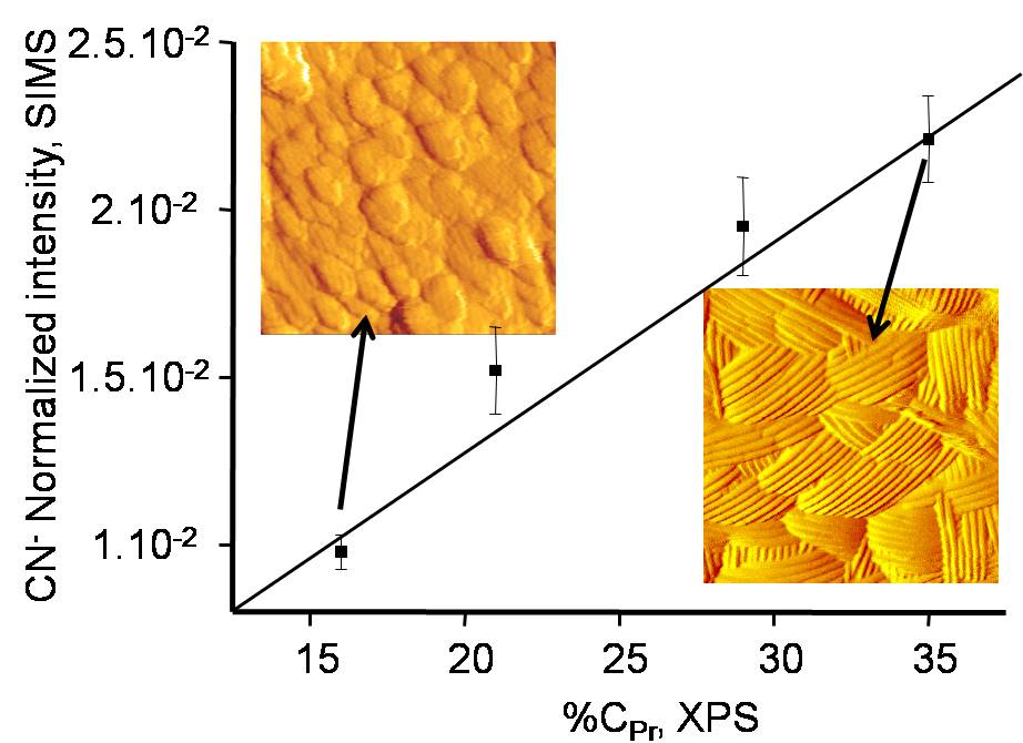
Combined use of atomic force microscopy, X-ray photoelectron spectroscopy and secondary ion mass spectrometry for cell surface analysis
Article accepté dans Langmuir IF=4.009


Simon Scheuring heads a small dynamic research team that is interested in "the structure and assembly of membrane proteins in native membranes studied by atomic force microscopy". This site provides contact address, references of publications, an image gallery, and a download section where you have access to CV and university degree reports.

Discussion sur les enjeux et risques des nanotechnologies suite à la projection du film le silence des nanos de julien Colin :
2 phrases à retenir et sur lesquelles réfléchir :
- si la réponse est les nanotechnologies, alors qu'elle était la question ?
- y avait-il un comité d'éthique pour empecher Cromagnon de fabriquer une massue ?
